
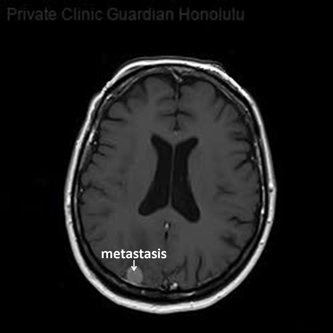
In T1-weighted magnetic resonance imaging (MRI) with contrast agent, an occipital cerebral metastasis is shown on the right side of the patient (note R = right) in layers 18 to 24. Cancer cells break away from the original (primary) tumor, travel through the blood or lymph system, and form a new tumor, then called metastases, in other organs or tissues (here in the brain) of the body.
© Wolfgang Schramm (2008)
• Homepage
• Zu deutscher Sprache wechseln