
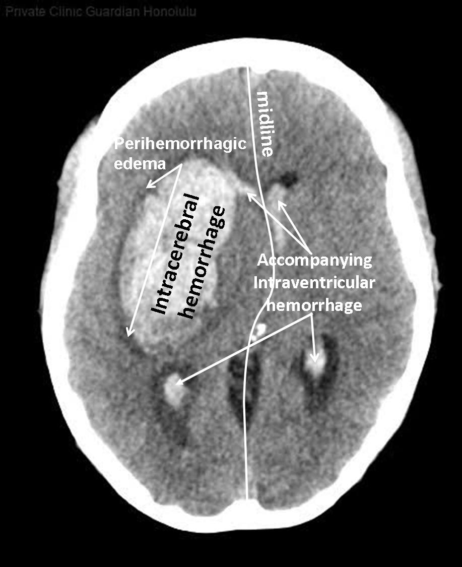
An intracerebral hemorrhage, or intraparenchymal cerebral hemorrhage, is a subset of an intracranial hemorrhage. In the layers 11 to 33 on the right side (note R = right) of this computed tomography (CT) without contrast agent there is an intracerebral hemorrhage (ICB) caused by arerial hypertension. An ICB is a bleeding into the brain tissue. This ICB (clearly visible in layers 23 to 25) has broken into the right anterior horn (= intraventricular hemorrhage). The fresh blood can be recognized on CT scans because this blood appears brighter than tissue. A perihemorrhagic edema is already evident (darker in the CCT), indicating that the bleeding event was already a few hours earlier. The midline shift to the contralateral side is pronounced, so there is an indication for surgical evacuation. Intracranial hemorrhage is common and is caused by diverse pathology, including trauma, hypertension, cerebral amyloid angiopathy, hemorrhagic conversion of ischemic infarction, cerebral aneurysms, cerebral arteriovenous malformations, dural arteriovenous fistula, vasculitis, and venous sinus thrombosis, bleeding into brain metastases or brain tumors among other causes.
© Wolfgang Schramm (2008)
• Homepage
• Zu deutscher Sprache wechseln