
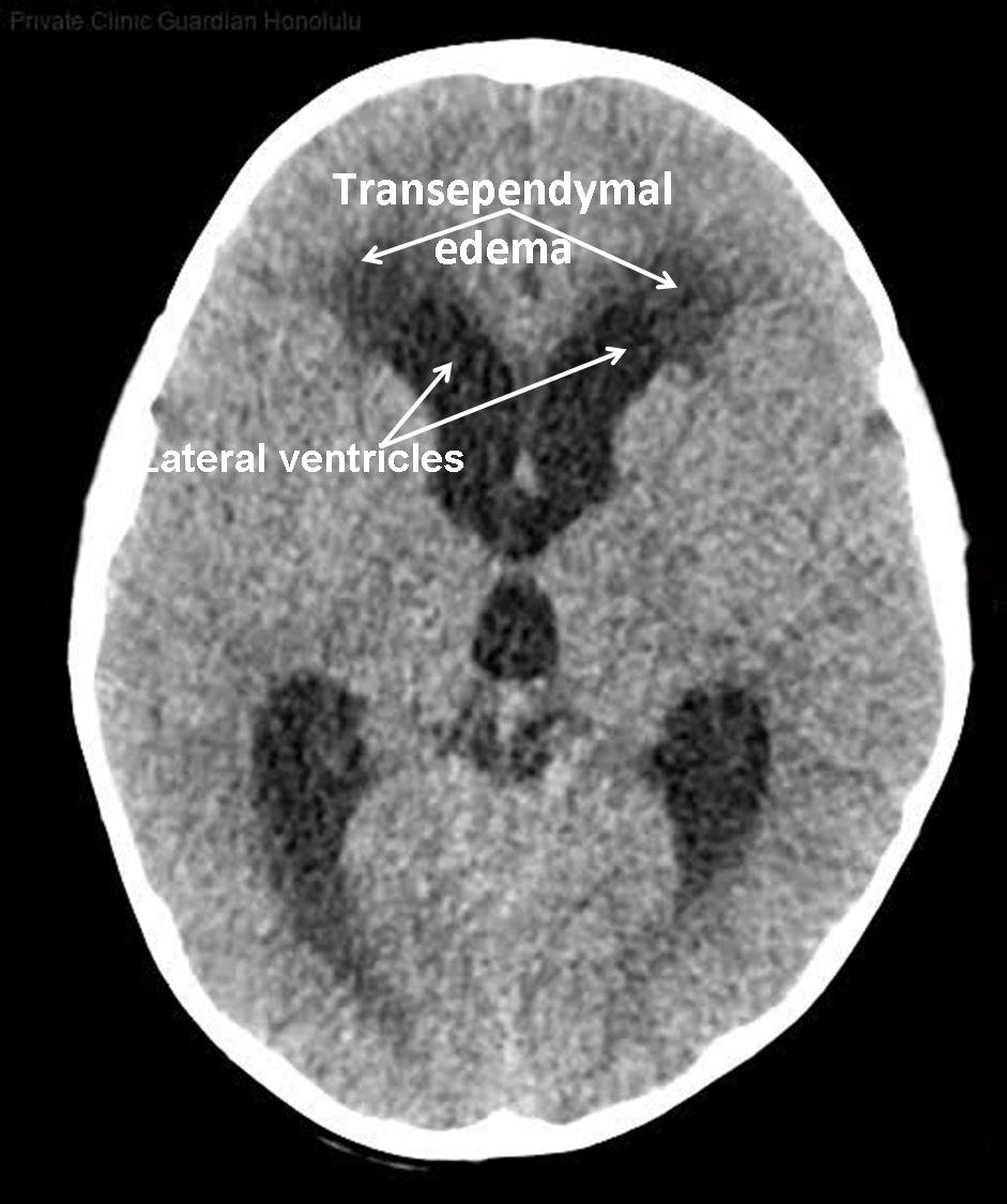
In layers 30 to 41 of this computed tomography (CT) of a 5-year-old child, there is an ependymoma with cystic and solid parts. Ependymomas are usually slow-growing tumors of the central nervous system that have histomorphologically similarities with ependymal cells. As a consequence of this tumor, a hydrocephalus with massively enlarged supratentorial fluid spaces and signs of a transependymal edema can be seen. The basal cisterns are clearly narrowed by the relocated 3rd ventricle, the sulci of the cerebral hemispheres have already passed and only the insular cistern is marked with a thin liquor marker.
© Wolfgang Schramm (2008)
• Homepage
• Zu deutscher Sprache wechseln