
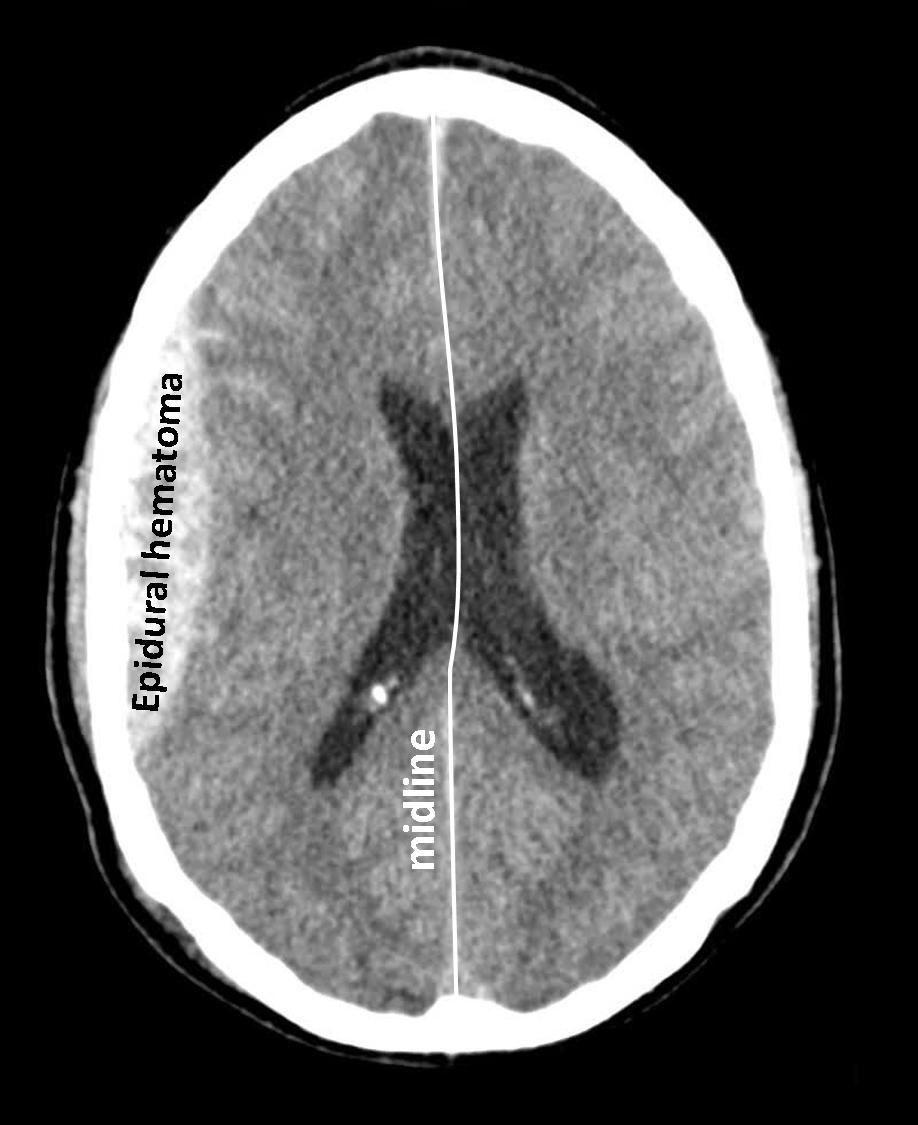
In cranial computed tomography, an epidural hematoma appears in layers 9 to 32 on the right side of the patient (note R = right). The hematoma already causes a slight shift of the midline to the contralateral side. In addition, there are several contusions bleeding. An epidural hematoma is a hemorrhage into the space between the skull bones and dura mater usually as a result of a rupture of the middle meningeal artery.
© Wolfgang Schramm (2008)
• Homepage
• Zu deutscher Sprache wechseln